
Apoorva Healthcare
A unit of Vittals Medicare Pvt Ltd

Apoorva Healthcare
A unit of Vittals Medicare Pvt Ltd


Radiology, also called diagnostic imaging, is a series of different tests that take pictures or images of various parts of the body. Many of these tests are unique in that they allow doctors to see inside the body. A number of different imaging exams can be used to provide this view, including X-ray, MRI, ultrasound, CT scan and PET scan.
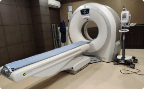
A computed Tomography (CT) scan uses x-ray in conjunction with computer algorithms to view the body's organs and tissues.This type ofscan can detect more subtle attenuations of x-rays images with the aid of a computer to generate cross sectional views of the internal organs and structures of the body.

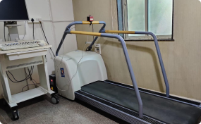
Heart health is an important factor in longevity of patient life. A treadmill test or stress test involves monitoring the patient’s heart rhythms to detect any abnormality as the patient walks on a treadmill overseen by a technician who increases the incline and speed on regular intervals so as to record the response of the patient’s heart. It is recommended for those who experience cardiac symptoms, most commonly chest pain.

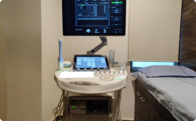
Ultrasonography uses high frequency sound waves to visualize soft tissue structures in the body. No ionizing radiation is involved and for this reason in considered safe to use in obstetrical imaging. Fetal anatomic development can be thoroughly evaluated allowing early diagnosis of many fetal anomalies.

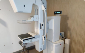
Mammography is a specific type of imaging that uses a low-dose x-ray system to examine breasts. A mammography exam called a mammogram, is used to aid early detection and diagnosis of breast diseases and cancers in women. An x-ray is a noninvasive medical test that helps physicians diagnose and treat medical conditions.

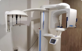
An OPG X-ray also known as Orthopantomagram is an instrument commonly used by dental doctors to understand the positioning, size, number and growth of all of the patient’s teeth. As an OPG provides the dental doctor a panoramic view of the patient’s teeth which covers multiple angles of the teeth in the upper jaw (maxilla) and lower jaw (mandible), it helps the dental doctor to provide an ideal treatment plan that covers orthodontic issues, development issues etc.

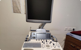
2D Echocardiography is generally recommended when the doctor suspects any malfunctioning in the various parts of the patient’s heart. The resulting image which is created by sound waves can help a cardiologist to understand potential cardiac risk factors that the patient might have including blocks, blood clots, tumours or any other such abnormalities through a non-invasive procedure.

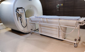
MMRI, or magnetic resonance imaging, is a means of “seeing” inside of the body in order for doctors to find certain diseases or abnormal conditions. MRI does not rely on the type of radiation (i.e. ionizing radiation) used for an x-ray or computed tomography (CT) scan.

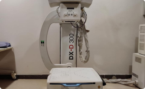
An X-ray is a quick, painless test that produces images of the body’s organs and structures but is predominantly use to produce images of normal and abnormal bones. X-rays can determine whether a fracture or break has occurred in any bone of the body. X-ray beams pass through the body and these are absorbed in different amounts depending on the density of the material they pass through.






 Rohan C
Excellent service & staff. Would especially like to thank Mr. Tejas for the efforts he has put in to get my reports 👍🏽
“
 Sandeep Rawat
Great and quick service, very hygienic working are and staff, the reports are accurate and get it ina timely manner. Would recommend Apoorva Diagnostic to all
“
 Nitesh Ranjit
Excellent time service provided !
“
 Deepshikha Choudhary
Achyut Sankhe we really appreciate your work. Thank you for all the quick responses always. With your strong follow up with your internal teams you have resolved our case within few hrs. This is much appreciated.... Thankyou and Great Work done by you.
“